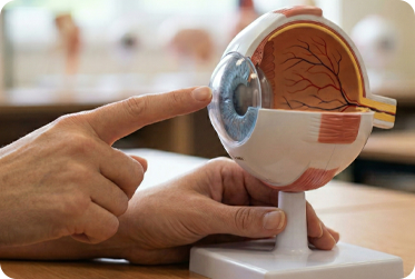
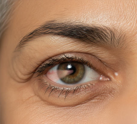
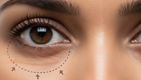
Symptoms of Corneal Problems
You should consult a specialist if you experience:
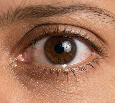
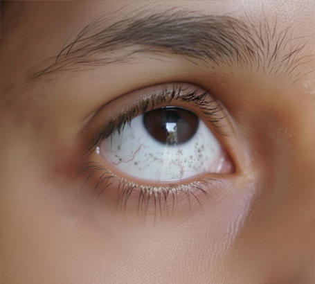
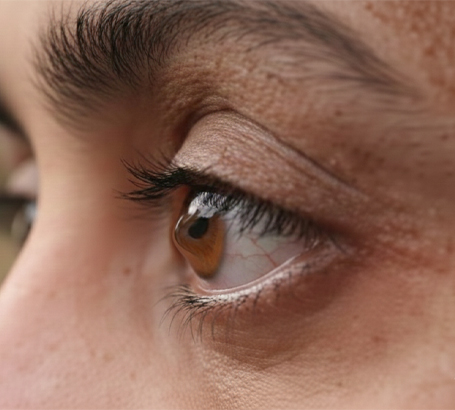
Blurred or distorted vision
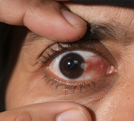
Eye pain or redness
Light sensitivity (photophobia)
Excessive tearing or discharge
Feeling of something in the eye
Early diagnosis helps prevent complications and vision loss.
Cornea Treatment Options
Treatment depends on the severity and type of corneal disease.
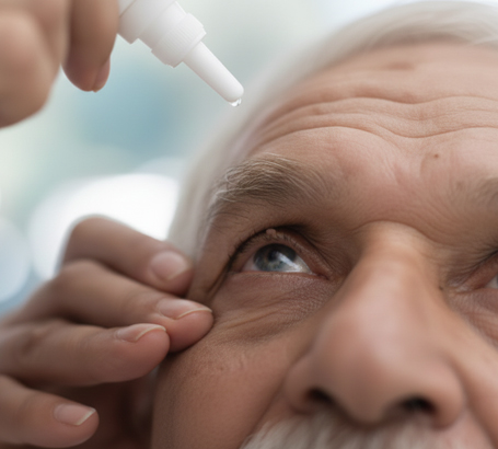
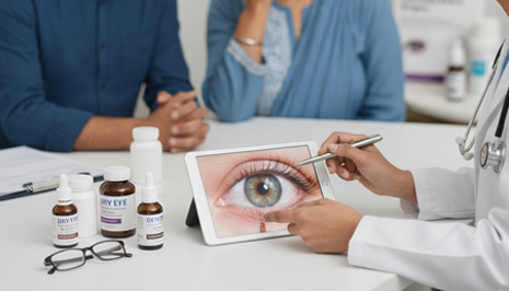
1. Medications (Eye Drops)
Used for infections, inflammation, and dry eye
Helps control early-stage conditions
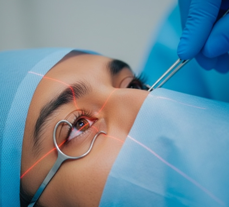
2. Corneal Cross-Linking (CXL)
3. Specialty Contact Lenses
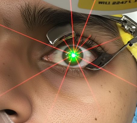
4. Laser Treatment (PRK/PTK)
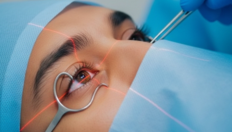
5. Corneal Transplant (Keratoplasty)
At ASG Eye Hospital, treatment is customized based on your eye condition and lifestyle needs.
Cornea Treatment Cost in India
The cost of cornea treatment varies depending on the condition and procedure required.
Factors Affecting Cost:
Type of disease (infection, keratoconus, transplant)
Treatment method (medication, laser, surgery)
Technology used
Hospital location
Estimated Cost:
Eye Drops / Medication: ₹1,000 – ₹5,000
Corneal Cross-Linking (CXL): ₹25,000 – ₹50,000
Laser Treatment: ₹20,000 – ₹60,000
Corneal Transplant: ₹80,000 – ₹2,50,000
We ensure affordable and transparent pricing with high-quality care.
Pre & Post Treatment Care
Proper care helps improve recovery and long-term results.
Before Treatment:
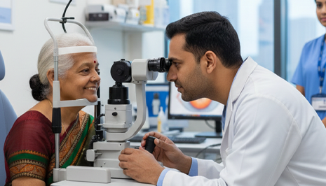
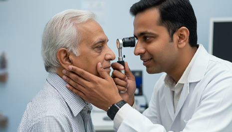
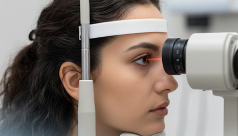
Detailed corneal examination
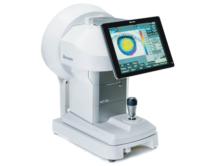
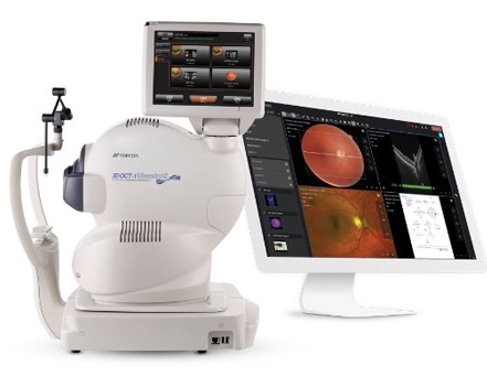
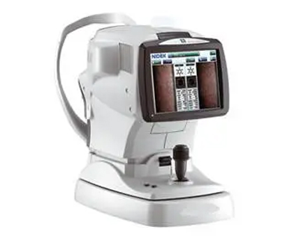
Corneal topography and imaging
Diagnosis and treatment planning
After Treatment:
Use prescribed medications regularly
Avoid touching or rubbing the eyes
Protect eyes from dust and infection
Attend follow-up visits
Most patients experience significant improvement with timely treatment.
Why Choose ASG Eye Hospital for Cornea Treatment?
Experienced cornea specialists
Advanced diagnostic tools (Topography, OCT)
Latest treatment techniques including transplant
High success rate in corneal procedures
Personalized patient care
ASG Eye Hospital is a trusted center for comprehensive cornea care in India.
Looking for Cornea Treatment Near Me?
If you are searching for cornea specialist near me or cornea treatment near me, ASG Eye Hospital provides expert care across multiple locations in India.
Book your consultation today and restore clear, healthy vision with expert cornea care.