A sudden vision issue in one eye may be written off as tiredness or a change in prescription. When it comes with pain on eye movement, faded colors, or a central dark spot, the nerve which transmits info from the eye to the brain may be affected. Optic neuritis needs timely assessment of such symptoms as many eye and neurological conditions may present similarly.
At ASG Eye Care we evaluate for signs of optic neuritis symptoms during a thorough eye exam which may be followed by a neurological review. This is an educational resource; should you experience or sudden onset of vision loss that is unexplained see your health care provider immediately.
What Is Optic Neuritis?
The optic nerve is made up of nerve fibers which carry electrical signals from the retina to the brain where they are interpreted as sight.Optic neuritis in many cases what we see is that the immune system attacks this area causing inflammation which in turn interferes with that pathway. Also in many instances the immune system affects myelin, the protective material which surrounds the nerve fibers and in doing so it slows the transmission of visual information.
The optic nerve is made up of nerve fibers which carry electrical signals from the retina to the brain where they are interpreted as sight. In many cases what we see is that the immune system attacks this area causing optic nerve inflammation which in turn interferes with that pathway. Also in many instances the immune system affects myelin, the protective material which surrounds the nerve fibers and in doing so it slows the transmission of visual information.
The course of optic neuritis is variable. Vision may go into mild blur or may drop off sharply over hours or days. Recovery is a function of the cause, subtype, and degree of the attack
Early Optic Neuritis Symptoms
The most optic neuritis symptoms are reduced vision and pain around or behind the eye which may present when looking to the side. At the onset the pain may be before the visual impairment or at the same time. Some report an ache or pressure as opposed to a sharp pain.
Color vision may fade out over time which in turn makes red objects appear gray or washed out. Also at reduced levels of light faces, text, steps, or items may become hard to tell apart which may not be very noticeable at first.
Other optic neuritis symptoms may present as a central blurred spot, missing areas in the visual field, flicker, or temporary deterioration which may occur with body temperature rise from fever, exercise, or a hot shower. Fever related dimness usually improves with cooling down but should still be reported during the exam.
| Visual change | How it may feel | Why assessment matters |
| Pain with eye movement | An ache behind the eye when looking sideways or upwards | It commonly accompanies inflammatory optic nerve disease, although other conditions can cause similar pain |
| Reduced clarity | Foggy, dim, blurred, or missing central vision | The speed and degree of loss help distinguish optic neuritis from retinal and vascular disorders |
| Altered colour vision | Red looks faded or colours seem washed out | Colour desaturation is one of the useful optic neuritis symptoms checked by the doctor |
| Reduced contrast | Faces, print, or steps appear less defined | Contrast may remain affected after sharpness begins to recover |
| Heat-related dimming | Sight becomes temporarily worse after exertion or heat | It can reflect disturbed signal conduction along an affected nerve |
These signs do not prove optic neuritis. Retinal disease, glaucoma, migraine, reduced blood supply, infection, and nerve compression can overlap, so examination is essential.
What Causes Optic Nerve Inflammation?
At times no cause is determined. Also in some patients optic nerve inflammation is related to an immune mediated or demyelinating disease. Multiple sclerosis is a known association and in some cases an episode which affects vision may be the first thing that brings a person in to the doctor for diagnosis. This is not to say that all people with optic neuritis have or will develop multiple sclerosis
Doctors also look at neuromyelitis optica spectrum disorder and myelin oligodendrocyte glycoprotein antibody associated disease. These conditions present a different picture of optic nerve inflammation which may involve both eyes causing severe and permanent loss which may be recurrent or of a larger section of the nerve. Proper identification of the subtype is key because it guides in the immediate care and long term prevention.
Systemic illnesses like sarcoidosis and lupus are related, also we see that viral, bacterial, or fungal infections may present. Age, immune status, recent health issues, exposure to illness, exam results, and neurological symptoms inform which tests to run
Not for every case of damaged optic neuritis do we see optic neuritis. That which causes reduction in blood flow, nutrition deficiency, toxic exposure, certain medicines, inherited disease, advanced glaucoma, or pressure from a mass may bring on optic neuropathy by a different route. Also we see that inflammation based treatment may not work or may in fact be unsafe when the cause is something other than infection.
Who Can Develop It?
Typical of young adults and what we see is that women report more frequently to us on this issue of optic neuritis but it does present at any age. We do see it in children and in the senior population which presents with similar issues of vision loss in these cases we have to do a very careful assessment for vascular or other causes.Bilateral affectation, large disc swelling, repeat attacks, lack of pain, or progressive decline may present an atypical picture. In which case specialist investigation for management of optic neuritis should be done instead of a standard treatment.
How Is Optic Neuritis Diagnosed?
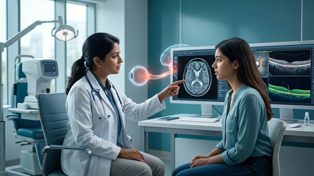
The doctor will ask if the vision change has happened, what kind of pain you have had, the speed at which it developed, and if there were any previous eye or neurological issues. In terms of testing we look at visual acuity, pupils’ reaction, color vision, contrast, eye movement, visual fields, the retina, and the optic disc. A relative afferent pupillary defect can show that visual info may be impaired in one eye as opposed to the other but it doesn’t tell us what the cause is.
Magnetic resonance imaging of the brain and orbits also performed with contrast when clinical indication presents, may present results of optic nerve inflammation and to also identify other changes in the central nervous system. MRI also used to rule out compression and to determine if a neurological evaluation is required.
Optical coherence tomography which looks at the retinal nerve-fibre and ganglion-cell layers and visual field testing which maps out areas of vision loss. As for the blood tests they may include for example infection, systemic inflammation, aquaporin-4 or MOG antibodies. Lumbar puncture is done only if the clinical picture indicates it does.
Assessment of these warning signs is more than a spectacle power check. Doctors have to determine if the nerve is affected, what the cause is, and what the course of action that may range from urgent therapy to long term neurological care will be.
Optic Neuritis Treatment
The right optic neuritis treatment is based on the degree of severity, which eyes are affected, what we think the subtype is, results of the MRI and blood work, and the patient’s health in general. Some typical attacks run their course without treatment, while in other cases we treat to speed recovery or in very severe or atypical presentations.
High dose corticosteroids are given to selected patients. From the Optic Neuritis Treatment Trial we see that IV steroids brought about faster recovery in typical cases but did not improve the final visual acuity which in fact had better results in the watchful waiting group. Also the study reported that low dose oral prednisone when used by itself raised the chance of a relapse which is why we stress that patients should never use left over steroid tabs or someone else’s prescription.
Modern in patient care for optic neuritis treatment we tailor the approach. The health professional will choose the drug, dose, route, duration, and also any taper. As with steroids which impact blood sugar, blood pressure, mood, sleep, and infection risk, we must balance benefits against the patient’s medical history.
| Treatment approach | When it may be considered | Purpose and limitation |
| Observation with close follow-up | Selected typical cases without concerning features | Vision may improve naturally, but follow-up must confirm that recovery follows the expected course |
| High-dose corticosteroids | Significant loss, bilateral disease, or a specialist-defined inflammatory pattern | May speed recovery; the regimen must be prescribed and may not improve final vision in typical cases |
| Plasma exchange | Severe attacks that respond poorly to steroids, especially in some antibody-mediated disorders | Removes circulating inflammatory antibodies and may be used as rescue therapy |
| Treatment of infection or systemic disease | When testing identifies a specific underlying cause | Addresses the driver of optic nerve inflammation rather than treating the eye alone |
| Long-term preventive therapy | Recurrent disease or a diagnosis such as NMOSD, MOG-associated disease, or multiple sclerosis | Aims to reduce further attacks and is planned with the relevant specialists |
When it comes to infections which may transpire steroids may be unsafe to use until the infection is also in treatment. Also for some antibody mediated attacks which may be more acute in their progression as recovery is not as certain, For these reasons, optic neuritis treatment should immediate action may be required which is why we present these as a clinical diagnosis.
Recovery and Follow-Up
In most cases what we see is an improvement which within a few weeks of the attack has taken place and may in some cases be still improving for months. Sharpness may return before color, contrast or depth perception does.
Recovery from optic neuritis is variable. In some it returns to nearly normal, in others there is a persistent central blur, reduced contrast, color desaturation, or visual fatigue. With repeat or atypical attacks of optic nerve inflammation there is a greater risk of permanent damage which may be a feature of severe attacks.
Follow up reports which detail the progress of optic neuritis symptoms. If vision continues to decline, does not see improvement at all, returns after some initial improvement, or which affects the second eye the diagnosis and management plan may require reevaluation.
When Is Urgent Eye Care Needed?
Immediate assessment is required for any new vision loss which develops over hours or days, which is in both eyes, or which presents with weakness, numbness, balance issues, bladder problems, persistent vomiting, severe headache, fever or any other neurological symptom.
A sudden painless loss of vision also requires emergency care which may not fit the usual pattern. Retinal and vascular crises are known to cause permanent damage if left untreated. Also note that marked redness, discharge, history of trauma, chemical burn, or severe surface pain may be signs of a different acute eye issue.
Do not operate vehicles when visibility is greatly reduced, and do not start on steroids without medical supervision. Early diagnosis is key to tell apart optic neuritis from other conditions which require a different treatment.
Living Through Recovery
Follow out the care plan which the ophthalmologist or neurologist has given. For better safety use large print, screen magnification, and glare reduced lighting. While avoiding over heating may help with temporary dimming it does not take the place of what is required in your recovery from optic neuritis. Report any new eye or nerve issues right away.
Frequently Asked Questions
1. Is optic neuritis always linked to multiple sclerosis?
No. Optic neuritis can be associated with some other immune disease, infection, systemic inflammation, or no cause is found. MRI and the full clinical picture inform neurological evaluation.
2. What are the first optic neuritis symptoms?
At the onset of optic neuritis symptoms you may see dim vision in one eye, pain during eye movement, faded colors, and a central or patchy area of reduced sight. sudden visual changes should be checked.
3. Can both eyes be affected?
Yes. Optic neuritis may present with involvement of both eyes in certain forms such as that seen in some cases of childhood, post-infectious, or antibody mediated disease. Prompt referral to a specialist is recommended at the first sign of bilateral visual change
4. Does optic nerve inflammation make the eye look red?
Usually in most cases. Optic nerve inflammation reports to be behind the eye which at the same time may appear normal. Redness is a different issue which presents its own set of conditions and should be looked at also in relation to what is seen visually.
5. Can spectacles correct the problem?
A new prescription will not treat present nerve related loss. In Optic neuritis symptoms appear because nerves are not transmitting information properly.
6. Is optic neuritis treatment always required?
In many cases not. For some patients observation is an option, while optic neuritis treatment is recommended in the case of severe, bilateral, recurrent, infectious, or autoimmune disease.
7. Are steroid tablets safe?
Steroids are to be used under specialist care as dose and route of administration is important. In terms of optic neuritis treatment safety also depends on management of diabetes, blood pressure, pregnancy, infection risk, and other health issues
8. Can the condition return?
Yes. Recurrence may present in one or both eyes. Repeated episodes of optic nerve inflammation are reasons for in depth evaluation and discussion of preventive options.
9. How soon should vision improve?
In many cases improvement is seen within weeks, but recovery may take months. Also if you have persistent or returning optic neuritis symptoms you should be re assessed.
10. Which specialist treats it?
An ophthalmologist assesses vision, at the same time a neuro-ophthalmologist or neurologist will do up imaging, antibody testing, and in depth care for complex optic neuritis treatment.
Conclusion
Optic neuritis is not the same as routine eye strain or a change in your glasses. Pain which comes with movement of the eye, colors that appear faded, reduced contrast, or quickly deteriorating vision may be a sign of the affected pathway between the eye and brain. Also because retinal, vascular, infectious, and neurological diseases can present in a similar way, it is better to get an early exam rather than to wait
ASG Eye Care also identifies causes of unexplained vision changes which at times may require referral to a specialist. Early diagnosis we use to determine the root cause, put in the best care plan, and we arrange for you to come back in for further evaluation of your visual and neurological health.