Blepharitis – Symptoms, Causes & Treatment
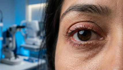
Blepharitis is a common eyelid inflammation that affects the edges of the eyelids where the eyelashes grow. It can cause redness, irritation, itching, burning sensation, crusting around the eyelids, watery eyes, and discomfort. Although blepharitis is usually not sight-threatening, untreated chronic inflammation may lead to dry eyes, recurrent eye infections, styes, and blurry vision. At ASG Eye Hospital, experienced eye specialists provide advanced diagnosis and personalized treatment for blepharitis and related eyelid disorders.
Types of Blepharitis
Anterior Blepharitis
Anterior blepharitis affects the outer front edge of the eyelid where eyelashes attach. It is often associated with bacteria or scalp dandruff.
Posterior Blepharitis
Posterior blepharitis occurs due to dysfunction of the meibomian glands located inside the eyelids. It is commonly linked with oily skin, rosacea, and dry eyes.
Mixed Blepharitis
Some patients may develop both anterior and posterior blepharitis simultaneously, causing persistent irritation and inflammation.
Symptoms of Blepharitis
Common symptoms of blepharitis include:
- Red and swollen eyelids
- Burning or itching sensation
- Crusting near eyelashes
- Watery eyes
- Dry eyes and irritation
- Sensitivity to light
- Sticky eyelids after waking up
- Frequent blinking
- Foreign body sensation in the eyes
- Blurred vision that improves after blinking
- Eyelash loss or misdirected eyelashes
If symptoms continue for a long period, consultation with an eye specialist is recommended.
Causes of Blepharitis
Blepharitis may develop due to several factors, including:
- Bacterial infection
- Blocked oil glands in eyelids
- Excessive dandruff
- Skin conditions such as rosacea or seborrheic dermatitis
- Dry eye syndrome
- Allergic reactions
- Poor eyelid hygiene
- Excessive screen exposure
- Contact lens irritation
- Environmental pollution and dust
Identifying the underlying cause is important for effective long-term treatment.
Risk Factors for Blepharitis
People with the following conditions may have a higher risk of developing blepharitis:
- Chronic dry eyes
- Oily skin
- Dandruff
- Rosacea
- Diabetes
- Allergies
- Poor eyelid hygiene
- Frequent eye makeup use
- Long screen usage
Complications of Untreated Blepharitis
Without proper treatment, blepharitis may lead to:
- Recurrent styes
- Chalazion formation
- Chronic dry eye disease
- Corneal irritation
- Eye infections
- Eyelash abnormalities
- Blurred vision
- Discomfort while wearing contact lenses
Early management helps reduce recurring symptoms and complications.
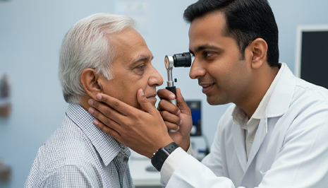
Diagnosis of Blepharitis
At ASG Eye Hospital, eye specialists perform detailed eye examinations to diagnose blepharitis accurately. Evaluation may include:
- Slit lamp eye examination
- Eyelid and eyelash assessment
- Tear film evaluation
- Meibomian gland assessment
- Dry eye testing
- Corneal examination
A proper diagnosis helps determine whether the condition is bacterial, inflammatory, or associated with dry eye disease.
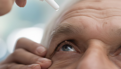
Blepharitis Treatment Options
Eyelid Hygiene
Regular eyelid cleaning is one of the most important steps in blepharitis management. Warm compresses and gentle eyelid cleansing help remove debris and improve oil gland function.
Warm Compress Therapy
Warm compresses help loosen crusts and improve blocked oil gland secretion, reducing irritation and inflammation.
Lubricating Eye Drops
Artificial tears and lubricating eye drops help relieve dryness and irritation associated with blepharitis.
Antibiotic Medication
In bacterial blepharitis, antibiotic eye drops or ointments may be prescribed to reduce infection and inflammation.
Anti-inflammatory Treatment
Some patients may require anti-inflammatory medications for chronic or severe inflammation.
Meibomian Gland Treatment
Advanced gland expression and dry eye management therapies may help improve eyelid oil gland function.
Lifestyle & Hygiene Guidance
Doctors may recommend reducing screen exposure, improving facial hygiene, controlling dandruff, and avoiding eye rubbing to prevent recurrence.
How to Prevent Blepharitis
You can reduce the risk of blepharitis by:
- Maintaining proper eyelid hygiene
- Removing eye makeup before sleeping
- Managing dandruff and skin conditions
- Avoiding touching or rubbing the eyes
- Taking breaks during screen use
- Keeping contact lenses clean
- Using prescribed lubricating eye drops
Why Choose ASG Eye Hospital for Blepharitis Treatment?
- Experienced eye specialists and oculoplasty experts
- Advanced diagnosis for eyelid disorders
- Personalized dry eye and blepharitis treatment
- Modern eye care technology
- Comprehensive eye examination facilities
- Trusted eye hospital network across India
- Patient-focused treatment approach