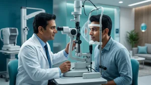

Clarity of vision is very much a function of the cornea, the transparent which is the front element of the eye. It allows light in and also plays a role in the light’s focus towards the retina. When corneal infection which in turn causes scarring, swelling out of shape and misshaping occurs, light is not passed through the cornea as it is supposed to. This results in a range of vision loss from mild blurring to total corneal blindness.
Corneal problems may show up as pain, redness, excessive tearing, sensitivity to light, or a visible white spot. Since some infections and injuries worsen quickly, prompt assessment matters. Treatment at an early stage can include medication, protective procedures, or surgery, and many patients can be managed with these approaches. When the damage is permanent and affects the central corneal transplant may replace the unhealthy tissue and help restore useful vision.
What Is Corneal Blindness?
Corneal blindness is a term for serious vision impairment which results from a cornea which has lost its transparency or normal shape. Usually the cornea is clear, smooth, and perfectly curved. It acts as a protective window which also provides a great deal of the eye’s focusing power. When its layers become cloudy or uneven, light entering the eye scatters rather than forming a sharp focus.
The term in question is a broad one which does not specify a single disease. It is a result of severe keratitis, corneal ulcers, trauma, chemical injury, inherited dystrophies or progressive thinning disorders. We see that what causes corneal blindness is variable and from that we determine which of the options of medicine, laser treatment, support care or surgery is the proper choice.
Damage at the edge of the cornea may have little effect on sight. In the center a dense scar may cause very serious visual impairment even when the retina and the optic nerve are healthy. That is why during exam the location, depth and activity of disease is determined.
Symptoms and Warning Signs
Symptoms present depending on the base health issue. In some cases which are gradual in nature we see the onset of glare, halos, distorted vision or a frequent need for change in prescription of glasses. With infection and injury we see sudden pain, redness and a quick decline in vision. Also we note that other symptoms include cloudy vision, excessive tearing, discharge, light sensitivity, a foreign body sensation and difficulty keeping the eye open.
A corneal ulcer will present a small grey or white spot, but some may be only apparent with the use of a slit lamp. Severe pain from contact lens wear, an agricultural accident, or out of date water needs to be checked out right away. The CDC reports that microbial keratitis is caused by bacteria, fungi, viruses or parasites and in serious cases may cause blindness or a corneal transplant.
| Presenting Signs and Symptoms | Possible concern | Recommended response |
| Sudden pain, redness and blurred vision | Infection, ulcer or abrasion | Seek urgent ophthalmic assessment |
| White or grey corneal spot | Ulcer, infiltrate or scar | Arrange a same-day eye examination |
| Marked light sensitivity and watering | Corneal inflammation or infection | Avoid self-medication and see an eye doctor |
| Gradually distorted vision | Keratoconus or corneal irregularity | Book a comprehensive corneal evaluation |
| Cloudiness after eye surgery | Swelling or another complication | Contact the treating surgeon promptly |
| Chemical exposure | Surface burn with risk of rapid damage | Rinse immediately and obtain emergency care |
Major Causes of Corneal Blindness
The causes of corneal blindness are many and vary by age, occupation, general health and access to prompt ophthalmic care. Corneal infection which also goes by the term infectious keratitis is a main cause. Bacteria, fungi, viruses and parasites may enter via a broken skin. Risk goes up after an eye injury, improper contact lens care or exposure to contaminated water. If left untreated infection may cause permanent central scarring.
Eye injuries are also a great issue. Metal particles, glass, thorns, dust and plant material which get into the eye can cause a scratch or penetration of the cornea. In agriculture this is very important as organic material may introduce fungi. Chemical burns from acids, alkalis or cleaning products may damage the eye within minutes. Timely irrigation and professional care can reduce the risk of permanent visual loss.
Keratoconus is a disorder which causes the cornea to gradually thin and bulge. In the early stages the disease may be managed with the use of glasses or special contact lenses and in some cases progression may be retarded with cross-linking. As the disease progresses to more advanced stages may include surgical options for corneal disease treatment.
Inherited corneal dystrophies may present when there is accumulation of abnormal substances in specific layers. Also, autoimmune inflammation, dry eye to a severe degree, eyelid issues, and problems from past surgery may also play a role as causes of corneal blindness.
Severe vitamin A deficiency which in turn causes dryness, ulcers and tissue death especially in at risk children. It is also a causes of corneal blindness. WHO reports xerophthalmia to be a range which from night blindness may progress to blind causing corneal ulceration and necrosis. Infection may cause scarring of the eyelid which in turn makes the eyelashes turn in and rub against the cornea. World wide it is a causes of corneal blindness of the infectious varieties. WHO in October 2024 declared India to have eliminated trachoma as a public health issue but we must still do better in terms of awareness and surveillance. on Diagnosis of Trachoma.
Diagnosis starts with a full history. An ophthalmologist may put questions to you on when the symptoms began, past trauma, contact lens use, past surgeries, chemical exposure and family history. Visual acuity testing which is also done to see how much sight has been affected.
A slit lamp provides a detailed look at the surface and into the deeper layers of the eye. Fluorescein dye can identify scratches, ulcers or areas which are missing epithelium. Corneal topography and tomography which map the shape and thickness is very useful for keratoconus. Pachymetry measures thickness while speculum microscopy looks at the endothelial cells which in turn help keep the cornea clear.
When infection is present, we may collect a small tissue sample for lab testing. Determining the cause will in turn guide us to the corneal disease treatment. Also we may check eye pressure, the lens, retina and optic nerve to assess how much vision will return post treatment of the cornea.
Treatment Options
Treatment is based on the cause, depth, location and rate of progression. In the case of infectious keratitis we require intensive use of appropriate antibiotics. The medication may be antibacterial, antifungal, antiviral or anti-parasitic. Steroids should not be used without an ophthalmologist as they may aggravate some infections.
Minor abrasions will heal with the use of lubricating ointments, protective measures and time but for more serious injuries more in depth monitoring is necessary. In case of a dry eye which has affected the surface of the eye treatments include preservative free lubricants, anti-inflammatory drugs, or interventions to improve tear production. Keratoconus is treated with special contact lenses or cross linking, in more advanced stages a corneal transplant may be required.
| Advanced corneal conditions. | Common approach | Main purpose |
| Bacterial, fungal, viral or parasitic keratitis | Targeted antimicrobial medicines and close review | Stop infection and limit scarring |
| Abrasion or foreign body | Removal when required, lubrication and protective care | Support healing and prevent infection |
| Keratoconus | Glasses, specialised lenses, cross-linking or surgery | Improve focus and slow progression |
| Superficial scar or dystrophy | Laser treatment in selected cases | Remove abnormal superficial tissue |
| Endothelial failure | Partial-thickness transplantation | Replace the damaged inner layer |
| Full-thickness scar or structural damage | Penetrating keratoplasty | Replace the entire damaged cornea |
When Is a Corneal Transplant Required?
A corneal transplant is recommended for permanent cloudiness, very thin cornea, severe distortion or one that won’t heal despite appropriate treatment. In surgery the damaged area is replaced with healthy donor tissue. Full thickness surgery is Penetrating Keratoplasty which replaces the entire thickness of the cornea, while partial thickness procedures only affect the affected layers. Also noted preservation of surrounding healthy tissue. Deep anterior lamellar keratoplasty is performed when the front and middle layers are affected but the inner layer is still healthy. DSEK or DMEK will replace the diseased endothelial layer in which there is continuous swelling. The choice of which procedure to do is based on the exam results and health of the rest of the eye.
Surgery does not guarantee perfect vision right away. Healing can take weeks or months, and some people may still need glasses or contact lenses. The prescribed drops help limit inflammation and reduce the risk of rejection. After a corneal transplant, redness, pain, light sensitivity, or newly cloudy vision requires immediate contact with the treating ophthalmologist. Early Protection
Not all cases are preventable which is true for inherited diseases in particular but we see that there is a range of causes of corneal blindness which may be reduced. Use of protective wear is recommended during farming, construction, grinding, welding and chemical handling. In the event of eye injury do not rub the eye and seek professional care for any foreign object which has embedded itself.
Contact lenses require that you follow the care instructions we give you exactly. Do not use tap water to clean them, do not share them and do not wear them past the time we have prescribed. Also avoid wearing while sleeping or swimming which increases the chance of infection. Good practice in lens care is a step to avoid severe infection and later corneal disease treatment which may require extensive treatment. Also note of adequate nutrition and prompt assessment of eye redness, injury or a white corneal spot. People with diabetes, autoimmune disease, chronic dry eye or past corneal surgery may require regular exams. Early identification of keratoconus, dystrophy or endothelial weakness can avert delayed care and reduce the chance of permanent scarring.
Eye Donation and Corneal Care in India
Donor tissue provides many people with treatable corneal opacity the chance to restore useful vision. The National Program for Control of Blindness and Visual Impairment supports eye banking, collection, training and transplant services. A Government of India report from March 2026 said that we have 396 functional eye banks and 69,848 corneas collected during 2024-25. Donor tissue is assessed for suitability.Tissue not appropriate for use in optical surgery still may be used for therapy, training or research. Cornea donation is possible at time of death at home or in hospital when an authorized eye bank is notified right away. Families should only work with recognized services and to follow official guidance.
Emergency evaluation is required for sudden sight loss, severe pain, redness, a white spot, chemical exposure, penetrating injury or increasing light sensitivity. A contact lens wearer with pain that worsens after lens removal should also present to the emergency department. Delay may allow an ulcer to deepen and increase the risk of permanent visual loss.
Home remedies, leftover drops and over the counter steroid combinations may put off symptoms while disease progresses. A slit lamp exam and proper corneal disease treatment can mean the difference between full recovery with medications and eventually requiring surgery. A personal corneal disease treatment plan should be reviewed as the eye heals.
Conclusion
Corneal blindness is a large problem but in many cases it is preventable or treatable if only we recognize the warning signs early. Infection, trauma, keratoconus, inherited diseases, nutritional deficiency and complications of surgery are key causes of corneal blindness. Pain, redness, light sensitivity, a white spot or sudden blurring should never be ignored.
Early identification of corneal disease treatment allows us to put in place like the use of drugs and performing laser procedures and cross-linking which will preclude the development of scarring. If the disease has progressed, we may have to do a partial or full thickness corneal transplant that will give a chance at better vision. At ASG Eye Care we do a very detailed evaluation of the cornea that will point out what the issue is, which layer is affected and from there will come up with the best approach.
Frequently Asked Questions
1. Is Corneal Blindness Permanent?
Corneal blindness is a variable condition which may present temporarily or permanently based on the cause and the extent of scarring. With early intervention, an active infection or swelling may resolve; but a dense opacity at the center of the cornea may require surgical intervention.
2. What Are the Common Causes?
The causes of Corneal blindness can result from infections, trauma, chemical burns, keratoconus, inherited dystrophies, severe dry eye, vitamin A deficiency, or complications following surgery.
3. Can Medicines Restore Clear Sight?
Medicines do treat infection, inflammation or surface disease but they don’t always get rid of a deep seated scar. The ophthalmologist chooses corneal disease treatment once the affected layer is determined.
4. Is Transplant Surgery Painful?
A corneal transplant is carried out under anaesthesia, which controls pain during the operation. Some mild irritation can occur while the eye heals; prescribed medicines should be taken exactly as directed.
5. How Long Does Recovery Take?
Recovery time is a variable which technique is used. Some partial thickness procedures see better results sooner than full thickness surgeries, but complete visual rehabilitation may still take many months. Continued corneal disease treatment and follow-up remain important.
6. Can Contact Lenses Cause Serious Vision Loss?
Contact lenses which in large part are safe when used as directed, do have risks which present themselves with poor hygiene, overnight wear and water exposure which promote microbial keratitis. Severe infection is a preventable cause of corneal blindness.
7. When Should I See a Cornea Specialist?
Seek early care for pain, redness, sensitivity to light, blurred vision, a white spot, injury or sudden visual change. Timely assessment may prevent corneal blindness and reduce the chance of needing a corneal transplant.