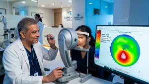
Keratoconus is a progressive condition which turns clear vision into a daily challenge. Signs may include double road signs, light glare from lamps, and very variable spectacle power. The disease develops when the normally round cornea thins and begins to bulge into an irregular cone like shape. At the point of diagnosis that this change is still occurring Corneal Cross-Linking may be recommended to strengthen the cornea and stop the progress.
This procedure plays an important yet defined role. It is a disease stabilizing Keratoconus treatment which does not promise perfect vision or freedom from glasses. That is to say that what we know of the process of CXL Surgery, what it does well, and what to expect during Keratoconus Recovery may help patients in their discussion with ophthalmologists.
Understanding Keratoconus
The cornea which is the front transparent structure of the eye also plays key role in light focus. In keratoconus this structure becomes gradually thinner and taller. This irregular shape causes blurred vision, ghost images, glare, light sensitivity, astigmatism, poor night vision and frequent prescription changes.
The disease does not affect all the same. In terms of diagnosis ophthalmologists use refraction, slit-lamp exam, corneal thickness measure, topography or tomography, and they also look to see how it has done in the past via early records to determine if the cornea is stable. We do2 not perform Corneal Cross-Linking Therapy at the first sign of Keratoconus we wait until we see progression which is confirmed by test results.
What Is Corneal Cross-Linking?
Corneal Cross-Linking is a low impact procedure which uses riboflavin, or vitamin B2, in combination with controlled ultraviolet-A light. What happens is that the treatment forms additional bonds between collagen fibers in the cornea which in turn makes the tissue stronger and more resistant to further change. The National Eye Institute reports this as a method to fortify the cornea and within FDA prescription info for a riboflavin based system it is put forth for use in progressive keratoconus.
Glasses and contact lenses improve how light is transmitted, but they do not reverse the structural damage of the cornea in which case they do not serve the same function. This is the primary role of this Keratoconus Treatment. It is to slow or stop progress of the disease in order to preserve present vision and options for the future.
When May the Procedure Be Recommended?
An ophthalmologist will bring up Corneal Cross-Linking as a treatment option which is supported by scan results that show an increase in corneal steepness, thinning, irregular astigmatism which is getting worse or changes in refraction which are repeated. Also included in the decision are age of the patient, corneal thickness, present scarring, visual requirements and health of the eye surface.
Timing is of the essence as a very thin or very scarred cornea may present limited options for treatment. In younger patients we see that progression may be faster at times which is why they require very close watch, but age in and of itself does not determine treatment. A personalized Keratoconus Treatment plan should be based on measurable change and a complete corneal assessment.
How CXL Surgery Is Performed
Before performing CXL surgery we perform a very detailed map of the eye and we measure the corneal thickness. Also at this time we may ask patients to stop wear of contact lenses prior to testing as they can temporarily change the corneal shape. We also go over past eye issues, allergies, present medications and any conditions which may affect the healing process.
In the traditional epithelium off approach which we use it is that we apply an anesthetic to the eye which in turn numbs it and we remove the thin surface layer of the cornea. We then apply riboflavin drops to the eye which in turn are taken up by the corneal tissue. The eye is put under a controlled dose of ultraviolet light. For a labeled FDA approved system which we use this includes corneal epithelial removal, repeated riboflavin application and measured doses of UV A light.
At the completion of Corneal Cross-Linking a band in the form of a contact lens is usually put in place which stays until the surface has healed. The ophthalmologist will give you a prescription for antibiotics, anti-inflammatories and lubricating drops. We ask that you follow that exact schedule and do not add in any over the counter eye drops.
Epi-Off and Epi-On Techniques
In epi-off CXL Surgery the surface epithel which is removed which in turn allows riboflavin to access the deeper corneal tissue. This is a well established practice but what one does see in the post op period is a few days of discomfort, watering and light sensitivity.
In epi-on, which is also transepithelial treatment the surface layer stays the same. This may present less initial pain but what varies between protocols is riboflavin delivery methods, equipment, eligibility and evidence. That decision should be made by a cornea specialist which takes into account corneal thickness, the stage of progression and the type of treatment available. Also the FDA has approved an epithelium-on riboflavin based system for eligible patients at age 13 and up in the U.S.
| Comparison | Epi-off technique | Epi-on technique |
| Corneal surface | Epithelium is removed | Epithelium remains intact |
| Riboflavin entry | Direct access to the corneal stroma | Requires a method designed for an intact surface |
| Early experience | More discomfort while the surface heals | Early discomfort may be lower |
| Recovery | Surface healing usually takes several days | Surface recovery may be faster |
| Selection | Based on corneal measurements and clinical judgement | Based on eligibility, protocol and available technology |
Benefits of Corneal Cross-Linking
The main benefit of Corneal Cross-Linking is in the area of corneal stabilization. What it does is strengthen collagen fibers which in turn is supposed to reduce the progressive steepening and thinning. NHS eye hospitals report that cross linking is a procedure which may delay or stop the progress of keratoconus although at the same time it is reported that patients may still require glasses or contact lenses.
Stability which maintains present visual function and may decrease the chance that more in depth corneal procedures will be put into use. Some patients report gradual flattening or moderate visual improvement, but this is variable and should not be presented as a given result of Keratoconus treatment.
Corneal Cross-Linking also leaves your future vision correction options open. Once the cornea has healed and stabilized, we may reevaluate the use of glasses, rigid gas permeable lenses, hybrid lenses or scleral lenses. Cross linking strengthens the cornea; these devices improve focus. Also do not think that one replaces the other.
What the Treatment Cannot Promise
Corneal Cross-Linking is also not the same as LASIK nor is it performed just to remove your glasses. It is not what guarantees a perfect cornea, a constant prescription or specific visual acuity. That some patients may see no change in their refractive error is a given even if progression has been very well controlled.
So the success of CXL Surgery is determined through follow up scans and long term stability which is why we look at the patient’s vision beyond the first few weeks post surgery. Also temporary blur post treatment is not in and of itself an indication that the procedure has failed.
Who May Not Be Suitable?
Suitability is a result of a full evaluation. Active infection, severe ocular surface disease, extensive corneal scarring, inadequate thickness or poor healing capacity may play a role in whether and how Corneal Cross-Linking is done. Past herpetic eye disease, pregnancy and some general health conditions must also be brought to the ophthalmologist’s attention.
Patients must present to us their full medical and eye history which in turn will inform our decision as to what is best for them we do not rely on general online eligibility rules. What is best for each patient in terms of Keratoconus treatment is what we base our decision on which is a close look at the measurements and condition of each individual eye.
Preparing for Treatment
Before undergoing CXL surgery please follow these instructions regarding contact lens removal, medications, diet, and time of your arrival. Also do not wear eye make up or use creams around the eye on the day of the procedure and please arrange for a ride home as your vision will be blurred post surgery.
Prepare a sterile, dim setting for rest. Have at hand as directed medications and also plan to reduce screen time, driving, dusty work and strenuous activity. These simple steps may improve the early Keratoconus Recovery process and also reduce avoid which may cause irritation.
Keratoconus Recovery: What to Expect
In the first to third days of Keratoconus recovery from epi-off treatment patients usually report the most discomfort. We see symptoms like watering, a gritty sensation, redness, light sensitivity and blurred vision as the surface heals. The bandage lens is removed only once the doctor determines that healing is sufficient.
Vision may fluctuate for a while before it improves. Haze, glare or blur may persist for several weeks and the corneal shape may change gradually over months. NHS reports that the eye may be sore for a few days and at first vision may be hazy.
| Recovery stage | Common experience | Main care priority |
| First 24–72 hours | Watering, discomfort, light sensitivity and blur | Rest, use medicines correctly and never rub the eye |
| First week | Surface healing and improving comfort | Attend review visits and keep water, dust and makeup away |
| Following weeks | Fluctuating vision, glare or temporary haze | Continue prescribed drops and lubrication |
| Following months | Gradual stabilisation of refraction and corneal shape | Repeat scans and reassess vision correction |
| Long term | Monitoring for stability | Keep follow-up appointments even when vision feels unchanged |
A smooth Keratoconus recovery will depend on your post op care. Do not rub the affected eye, stay away from swimming and contact sports until you are given the go ahead. Back to work, driving and contact lenses should be based on how you are feeling, your vision and what your ophthalmologist says rather than any online timetable.
Medicines, Follow-Up and Warning Signs
After CXL Surgery we prescribe drops which will reduce the risk of infection, control inflammation and in turn support the healing process. Before use wash your hands and see that the bottle tip does not touch the eye or eyelashes. Also do not use anesthetic drops at home without specific medical direction as to do so repeatedly and without supervision may cause damage to the cornea.
During the course of Keratoconus recovery report to the hospital right away if pain which was at first going down now returns, redness increases, you see discharge, your eyelids become very swollen or vision drops suddenly. Infection, delayed surface healing, inflammation, haze, scarring and reduced vision are known to be possible complications although it may vary by person.
Vision After Treatment
Patients report at times that they still require glasses or specialty contact lenses post Corneal Cross-Linking. While a stable cornea is a goal which makes future visual rehabilitation more predictable it is also true that lens fit is delayed until the surface and refraction has settled. In the ongoing plan for Keratoconus Treatment we include evaluation of the non affected eye which may progress independently.
Long term care is a part of Keratoconus treatment. Also, patients must give up the habit of eye rubbing and report in to their health care provider any allergies which cause them to itch. At follow up topography or tomography the specialist will compare the cornea’s shape with that of early reports.
Why Timely Assessment Matters
Frequent changes in prescriptions, growth of ghost images, poor night vision or reduced clarity with new glasses are signs to get a detailed corneal assessment. For Corneal Cross-Linking treatment the right time is determined by progression of the disease and corneal measurements which may not be at the onset of symptoms.
At our ASG Eye Hospital we have cornea specialists that will assess the stage and type of keratoconus you have and which treatment option is best for you. This may be monitoring, specialty lenses, CXL surgery or another Keratoconus Treatment. Early assessment gives patients the chance to protect the cornea before it advances into more severe thinning or scarring.
Frequently Asked Questions
1. Is corneal cross-linking painful?
The eye is anesthetized during the procedure which is why usually what you don’t see is the sharp pain. Post epi-off treatment it is typical to experience a few days of discomfort, watering and light sensitivity. We prescribe medicine and the bandage contact lens to help out in the early stages of Keratoconus recovery.
2. Does the procedure cure keratoconus?
Corneal Cross-Linking is to stabilize the cornea not to cure the condition. It does not guarantee that no change will take place in the future which is why we still require repeat scans and long term follow up.
3. Can vision improve after treatment?
Some people see a slow improvement in vision or in the cornea’s shape, but our primary goal is prevention of progression. In a responsible approach to Keratoconus Treatment we put stability first and address vision correction once healing has taken place.
4. How long does recovery take?
Surface symptoms of Keratoconus may improve in a few days’ time but vision may fluctuate for weeks and corneal measurements may continue to change for months. The rate of Keratoconus recovery is a variable which includes eye health and individual healing.
5. Will contact lenses still be required?
Many patients are still in use of glasses, rigid lenses or scleral lenses post CXL Surgery which we note is a result of the procedure’s strength on the cornea but by and large it does not remove that irregular astigmatism. Refitting is at a wait until we see the level of stabilization.
6. Can both eyes be treated together?
That decision is based on the patient’s condition, vision in each eye, corneal thickness, which protocol is to be used and the surgeon’s approach. Some patients are treated one eye at a time which allows the first eye to heal before the second procedure.
7. Can keratoconus progress again?
Progress in disease may be seen in a few cases which is why we still do follow up after Corneal Cross-Linking. We do repeat scans which help the ophthalmologist to determine that the cornea is stable.
8. When should I call the hospital?
Seek immediate advice if you notice that pain, redness or swelling is getting worse, discharge has appeared, or your vision has dropped suddenly during Keratoconus recovery. Do not wait for your next routine visit or to treat it with over the counter eye drops.