Many people are shocked to find out that retinal holes may develop pain free. Also it is the case that some patients only report a retinal hole during a routine eye exam, while at other times they may see warning signs which over time they can no longer ignore.
As the retina is the component which captures light and sends visual signals to the brain even a small retinal hole is an issue which may affect vision if left untreated. What is of greatest worry with a retinal hole is not the hole itself but the issues which follow.
When a retinal hole is allowed in fluid under the retina it increases the risk of retinal detachment which is a serious issue that may cause permanent vision loss if left until late stage. It is important for patients to know the symptoms, causes and what treatments are available which in turn will help them to seek out timely care and in this way to protect their eyesight.
What Is a Retinal Hole?
A retinal hole is a little opening that develops within the retina, the very thin tissue at the back of the eye. The retina’s role is to convert light into signals which travel to the brain thus enabling us to see clearly.
When a retinal hole forms what we have is a weak point in this very delicate tissue. In many cases a retinal hole will appear as a part of the natural aging process. As the gel-like vitreous inside the eye shrinks with age it may pull at the retina and thus create a small opening. Also certain eye conditions, severe myopia (high minus power), past eye injuries, and family history play a role in increasing the risk of developing a retinal hole.
Although not all retinal holes go on to cause serious vision issues, close monitoring and timely intervention is usually required to prevent further damage.
Early Retinal Hole Symptoms
One issue with retinal holes is that symptoms present very individually. Some people report no symptoms at all, while others present with sudden visual disturbances. We see that patients report flashes of light which may come up in dim lighting or when they move their eyes rapidly. Also very common are new floaters that present as little dots, cobwebs or shadows that drift within the field of vision. In some cases the retinal hole may cause blurry vision or a feeling that a part of the visual field is missing. In any case of a sudden increase in floaters or flashes, that should not be ignored. Though these symptoms may not always indicate a retinal hole, it is best to see a retina specialist for an immediate check up to rule out more serious retinal issues.
Who Is at Risk of Developing a Retinal Hole?
The likelihood of developing a retinal hole increases with age, particularly after the age of 40. However, several other risk factors can contribute to its development.
| Risk Factor | How It Increases Risk |
| High Myopia | Increased stretching of the retina |
| Ageing | Vitreous gel shrinks and pulls on retina |
| Eye Trauma | Direct injury can damage retinal tissue |
| Previous Eye Surgery | May increase retinal stress in some cases |
| Family History | Genetic predisposition to retinal conditions |
| Retinal Disorders | Existing retinal weakness increases risk |
People with high myopia are particularly vulnerable because the retina tends to be thinner and more fragile. Regular retinal examinations are often recommended for these individuals.
How Is a Retinal Hole Diagnosed?
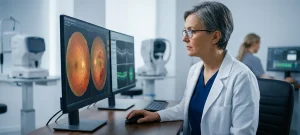
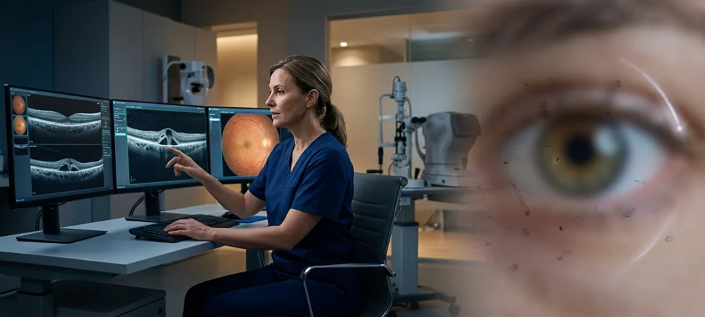
Diagnosis of a retinal hole is achieved through a full retinal exam. At the time of evaluation we which is to say that the ophthalmologist will dilate the pupils in order to get a very detailed view of the retina. Also at times special imaging tools will be used to determine the exact location and size of the retinal hole. We do a very in depth assessment which in turn helps to determine if the retinal hole is stable or if we in fact do need to do some sort of treatment in order to prevent it from progressing. Early diagnosis at this stage also plays a very large role in the preservation of vision.
When Does a Retinal Hole Require Treatment?
Not in all cases of retinal hole does intervention present as a must. Some small holes do in fact go for many years without issue. That said we do see to it that we treat when we note that the retinal hole is a player in the issue of retinal detachment. What we do in each case is look at a number of variables which include the retinal hole’s location, what other symptoms may be present, the patient’s health profile and also note the presence of fluid around the.affected area. This is why evaluation by a qualified retina specialist is extremely important.
Understanding Retinal Hole Laser Treatment
The most used intervention for symptomatic retinal holes is retinal laser hole treatment. This procedure uses concentrated laser energy to produce small burns around the affected area. What we see are laser marks which in turn form scar tissue that seals the retina to the base layers and which in turn reduces the risk of retinal detachment. Retinal hole laser treatment is an outpatient procedure which does not require hospital stay. Most patients go home right after the treatment. What we have here is a preventive measure. As opposed to improving present vision the procedure is to stop a retinal hole from turning into a more serious condition.
What Happens During Retinal Hole Laser Treatment?
During retinal tear laser treatment we use special eye drops which dilate the pupil and also numbing agent for the eye. The doctor then applies laser to the retinal hole with a special laser device. The procedure is usually for a few minutes. Most patients report retinal hole laser treatment to be only mild discomfort rather than pain. Vision may stay blurry for a short time after due to the dilating drops. In many cases retinal hole laser treatment does very well in creating a protective seal around the affected area and greatly reduces risk of retinal detachment.
Recovery After Retinal Hole Laser Treatment
Recovery from retinal hole laser treatment is usually a simple process. Post procedure patients may see some mild blurring, light sensitivity, or discomfort which is temporary. What you may notice are the laser scars that develop during retinal hole laser treatment which will take 1 to 2 weeks to properly heal at which point a strong seal is formed. While this is happening it is advised that patients follow what the doctor says and keep to the follow up schedule. Also of importance is the fact that a person who has had one retinal hole may go on to have more retinal changes in the future so we must remain vigilant and get checked out regularly.
Retinal Hole vs Retinal Detachment
Many patients confuse a retinal hole with retinal detachment, but they are not the same condition.
| Condition | Description | Urgency |
| Retinal Hole | Small opening in the retina | Requires evaluation and possible treatment |
| Retinal Tear | Larger break in retinal tissue | Higher risk of progression |
| Retinal Detachment | Retina separates from underlying tissue | Medical emergency |
A retinal hole can sometimes progress to retinal detachment if fluid passes beneath the retina. This is why early diagnosis and appropriate retinal hole laser treatment are so important.
How Can You Protect Your Vision?
The best way to care for your vision is through routine eye exams which we recommend especially for people with high myopia, diabetes, or a family history of retinal disease. Also if you see sudden flashes, floaters, or changes in your visual field seek out a retinal specialist right away.
We have seen great results in what modern treatment has to offer for patients with retinal conditions. We use procedures like retinal laser photocoagulation and retinal hole laser treatment which do a great job of preventing vision threatening complications when we catch them at the right time.
At ASG Eye Hospital our teams of experienced retina specialists use the best diagnostic technology and an evidence based approach to retinal disease treatment which we use to identify problems early and preserve vision when we can.
Conclusion
A retinal hole may not appear to be a big issue, yet they should be paid attention to. Symptoms which may present early as flashes of light, new floaters, a blur to vision, or a dark area within the visual field can be signs of a retinal hole. It is at the onset of these that you seek out an evaluation by a retina specialist and also, if required, have the issue treated with laser; which in turn can greatly lower the chance of retinal detachment and long term vision loss. Should you notice sudden changes in your vision the best thing you can do is to get in to see a professional right away.
Frequently Asked Questions
1. What are the symptoms of a retinal hole?
Flash and light symptoms are very common for a retinal hole as well as a sudden appearance of floaters, blurred vision and at times a shadow or curtain like effect in the field of vision.
2. Is retinal hole laser treatment painful?
For the most part patients report no pain at all during retinal hole laser treatment. Some may see minor discomfort or brief flares of light during the procedure.
3. Can a retinal hole heal on its own?
A retinal hole does not tend to close by itself. Your ophthalmologist will determine what is the best approach which may range from monitoring to retinal hole laser treatment.
4. How successful is retinal hole laser treatment?
Retina laser treatment for holes is very successful at preventing detachment if the procedure is done prior to major issues.
5. When should I see a retina specialist?
You should take it upon yourself to see a retina specialist as soon as you see flashes of light which are sudden, notice new floaters, have blurred vision, or see a dark shadow in your visual field.