
Retrobulbar neuritis is a condition which presents with inflammation of the optic nerve behind the eye. The optic nerve is responsible for taking visual information to the brain, thus damage in this area may come on very quickly in terms of vision. At first what we see is that patients report blurred vision, pain which comes and goes as they move the eye, reduced ability to see color, or a dark spot in the center of their vision. In retrobulbar neuritis the outer structure of the eye may look normal which in turn means that the issue is with the nerve tissue behind the eye which is inflamed.
This is the reason why the condition should not be ignored. Retrobulbar neuritis does improve with prompt care but also may be associated with multiple sclerosis, neuromyelitis optica, infections, or autoimmune disorders. At ASG Eye Care we aim for early diagnosis, vision protection, and the right treatment plan.
What Is Retrobulbar Neuritis?
Retrobulbar neuritis which is a type of optic neuritis has the affected element of the optic nerve located behind the eye. Also unlike some other eye diseases which present with obvious signs at first look, at primary evaluation the front of the nerve may appear normal. This may in some cases be a confusing picture although the symptoms are real and at large there may not be any redness or swelling.
In retrobulbar neuritis the primary issue is that of optic nerve inflammation. When the nerve is inflamed visual messages do not travel to the brain properly. As a result the patient may see dimly, have reduced color depth, see colors as brighter than they are or experience pain when the eye moves. As this affects the visual nerve pathway it is also classified as a type of eye nerve disease which requires careful evaluation.
Early Warning Signs
The issue is usually with one eye but in some cases both may be affected. Pain during eye movement is a key warning sign which we see as the inflamed nerve becomes sensitive to movement. Also many report that colors have faded out, especially red items, or that their vision appears gray and dull.
| Warning Sign | What It May Feel Like |
| Blurred or dim vision | One eye appears unclear, foggy, or darker |
| Eye pain on movement | Pain increases when looking side to side |
| Reduced colour brightness | Colours look washed out or less vivid |
| Central dark patch | A shadow or blur appears in the centre |
| Poor contrast | Reading or identifying objects becomes harder |
These optic neuritis symptoms should be checked early. Each episode of blurred vision may have a different cause which also includes that we out rule other vision loss causes like retinal disease, glaucoma, migraine related visual disturbance, stroke related vision problems, or severe dry eye.
Causes of Retrobulbar Neuritis
The causes vary between patients. In many young adults we see it as a result of an immune reaction which affects the sheath of the optic nerve. Also retrobulbar neuritis may be the first indicator of multiple sclerosis in some patients. As for other patients the cause may be infection, neuromyelitis optica, MOG antibody disease, autoimmune disease, vitamin deficiency, or exposure to certain toxins.
| Possible Cause | Why It Matters |
| Multiple sclerosis | May first appear with optic nerve-related vision loss |
| Neuromyelitis optica or MOG disease | Can cause severe or repeated attacks |
| Viral or bacterial infections | May trigger nerve inflammation |
| Autoimmune disease | The immune system may attack nerve tissue |
| Nutritional or toxic causes | Deficiency or toxin exposure may damage the nerve |
Because of the many vision loss causes diagnosis should not be delayed. Treatment of only the symptom without finding the cause can affect recovery. In some people retrobulbar neuritis is a single episode; in others it may return or point to a larger neurological issue.
How Doctors Diagnose It
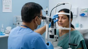
Diagnosis starts with a comprehensive eye exam. An ophthalmologist will test visual acuity, color vision, pupil response, eye movement, and the back of the eye. In retrobulbar neuritis the optic disc may at first appear normal so we often require other tests.
OCT is used to study the optic nerve and retinal nerve fiber layer. Visual field testing is done to identify blind spots or areas of reduced sensitivity. MRI may be advised to look for optic nerve inflammation, brain lesions, or signs linked with multiple sclerosis. Also we may do blood tests if we suspect infection, neuromyelitis optica, MOG disease, or an autoimmune disease. This approach also helps in confirmation of the diagnosis and in the elimination of other optic nerve diseases.
Treatment Options
Treatment is based on the degree of vision loss and the cause. In mild retrobulbar neuritis which may improve spontaneously we may just have a close watch and follow up. In moderate to severe cases doctors may put patients on high dose corticosteroids to reduce the inflammation and speed visual recovery.
Steroids will be given through IV or by mouth as per the case. If retrobulbar neuritis is a result of multiple sclerosis, neuromyelitis optica, infection or an autoimmune disease then treatment will go at that root cause. This may require a team up with a neurologist or another physician. Also it is important for patients not to self medicate with steroid tablets as improper treatment may worsen some infections or delay in getting to the right diagnosis.
Recovery and Long-Term Outlook
Recovery from optic neuritis is a very individual thing. Many see an improvement in a few weeks’ time which is great, but it may be that color vision, contrast and clarity which play up a bit more. Some people recover almost fully, others are left with some dimness or issues with contrast. We also must do follow up tests regularly as this condition can come back.
If optic neuritis symptoms return or if pain and vision loss worsen, urgent review is needed. Early treatment of optic nerve inflammation gives the best chance of protecting vision and reducing long-term damage.
Eye Care at ASG Eye Care
At ASG Eye Care we provide in depth assessment of our patients who present with what may be retrobulbar neuritis. Our full evaluation includes vision tests, nerve in the eye assessment, imaging studies and if required we also provide referral to a neurologist. As the signs of this condition may present very subtly, we do very thorough testing before we determine a course of treatment. We have a team of professionals that help patients to tell the difference between a common eye issue and a serious neurological condition.
Whether the concern is sudden blurred vision, pain with eye movement, repeated optic neuritis symptoms, or suspected nerve inflammation, timely consultation can make a meaningful difference.
Key Takeaway
Retrobulbar neuritis is not what one would term as common eye strain or transient blurring. It is a type of nerve inflammation which may present suddenly and at times is a symptom of a more serious health issue. If you are experiencing pain in the eye which gets worse with movement, a dulled color perception, a blurred center vision, or vision loss that comes out of the blue, early diagnosis may be key in preserving your sight.
Frequently Asked Questions
1 What is this condition?
Retrobulbar neuritis which is also referred to as optic neuritis is inflammation of the optic nerve at the back of the eye. This can present with blurred vision, pain which comes out with eye movement, reduced color vision, and central vision defects which may not be present when the eye is looked at from the outside.
2 What are common symptoms?
Common optic neuritis symptoms include pain while moving the eye, sudden onset of blurred vision, loss of color vision, poor depth perception and a dark/dim spot in the center of vision.
3 Can it cause permanent vision loss?
Most patients have partial or full recovery, but in some cases of severe or repeat retrobulbar neuritis there will be permanent visual changes. Risk factors for this are based on the cause, the degree of the issue, and when treatment is started.
4 Is it an emergency?
Sudden vision loss which also brings about pain in the eye is a medical emergency. This disease may require that we do eye tests, MRI, blood tests and also that we treat the base cause.