A sudden onset of floaters, repeated flashes of light or a dark curtain going over your vision is very alarming. These may be signs of a Retinal Detachment which is when the light sensitive retina detaches from the support structure. As the retina may lose function while it is detached, prompt examination is essential. In most cases Retinal Detachment Surgery is required to put the retina back in place, to repair the responsible tear and to protect what site is left. The National Eye Institute reports that a detached retina is a serious condition which requires prompt treatment.
Treatment is tailored. What is done depends on the cause, tear location, detached area, scar tissue, lens condition and if the macula has also detached. This guide details how Retinal Detachment Surgery is conducted, what to expect during the recovery period and which elements play a role in the outcome.
Understanding Retinal Detachment
The retina is a light sensitive layer of nerve tissue at the back of the eye. Most often Retinal Detachment comes about due to a retinal tear. Fluid gets in through the tear and accumulates under the retina which in turn detaches from the wall of the eye. Also there are other types which present when scar tissue pulls at the retina or when fluid accumulates without a tear. The National Eye Institute reports that these are known as rhegmatogenous, tractional and exudative detachments.
A medicine or glasses do not usually repair true Retinal Detachments. Laser or cold treatment may close a small tear but for large detachments a procedure which also repairs and puts the retina back in place is required. The goal of Retinal Detachment Surgery is to reattach the retina; how vision improves post surgery depends on how well it was doing before the detachment.
Why Early Treatment Matters
Timing is of the essence which the macula does for detail in our vision. While the macula is still attached which is what does the job of central vision, at that point in time urgent treatment may help to a great degree in preventing central sight from going. Once it has detached we still have Retinal Detachment Surgery which is important but the length of time and the degree of macular involvement may in fact limit what we see in terms of recovery. Flashes, floaters or that curtain-like shadow of detachment should not be waited out by the patient.
The warning signs which may not present pain at all. A sudden increase in floaters, new light flashes, blurred vision or a spreading shadow is a reason to go in for an emergency dilated retinal exam. MedlinePlus reports that delayed treatment may result in permanent vision loss.
Types of Retinal Detachment Surgery
The 3 main approaches are pneumatic retinopexy, Scleral Buckle Surgery and Vitrectomy Surgery. Laser photocoagulation or cryotherapy is also very often applied in these procedures for the purpose of causing a scar around the retinal tear. Per the National Eye Institute these are the primary surgical remedies that we have for a detached retina.
Pneumatic Retinopexy
Pneumatic retinopexy is used in certain, fairly simple cases of retinal detachment. The doctor injects a gas bubble into the eye which then the patient has to position their head in such a way that the bubble presses against the retinal tear. Then laser or cold treatment is applied to seal the break. This type of Retinal Detachment Surgery also requires very precise head positioning post procedure.
The gas bubble gradually disappears. Until the surgeon confirms that it has fully absorbed, the patient must not fly, travel to high altitude or receive nitrous-oxide anaesthesia. Changes in pressure can expand the bubble and dangerously raise pressure inside the eye. NHS guidance also warns patients with an intraocular gas bubble against flying until it is safe.
Scleral Buckle Surgery
During the Scleral Buckle Surgery the surgeon puts a silicone band or sponge on the white outer wall of the eye. The buckle which is applied there indents the eye wall towards the detached retina thus reducing traction at the tear. Also we use laser or cryotherapy to seal the break, and when needed fluid under the retina is drained.
Scleral Buckle Surgery may in some cases be performed on younger individuals, with which still have a natural clear lens, and detaches that are a result of certain peripheral breaks. The buckle is typically made permanent which in most cases is also not noticeable. As it does change the eye’s shape a new spectacle prescription may be given post repair.
After Scleral Buckle Surgery patients may see some redness, swelling, watering or discomfort. We do follow up checks on retinal position, eye pressure and healing. In some complex cases the buckle is used in conjunction with an internal procedure. Government and NHS resources present the buckle as an external implant which presses the eye wall into place at the retinal tear.
Vitrectomy Surgery
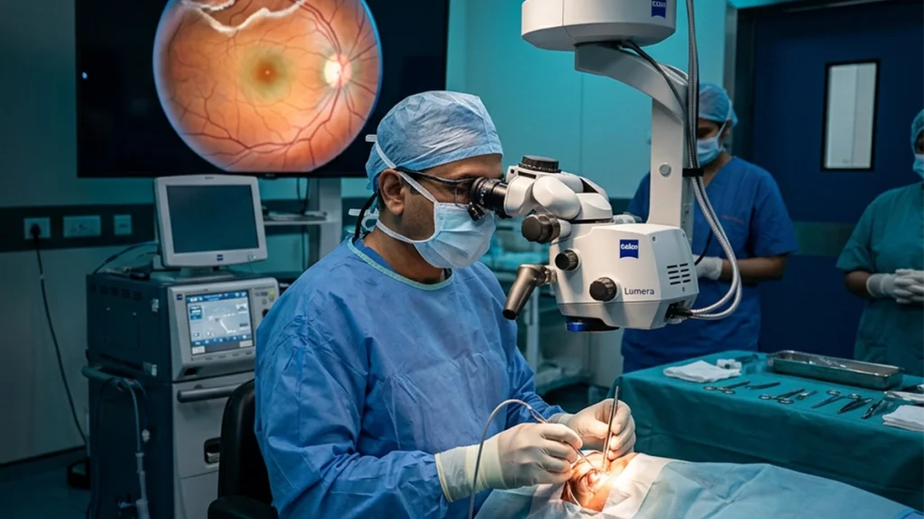
During the course of a vitrectomy surgery the doctor will make small incisions in the eye and remove the vitreous gel which may be tugging at the retina. The retina is flattened out, breaks in the retinal are repaired and the eye is filled with air, gas or silicone oil to hold the retina in place as it heals.
Vitrectomy surgery is a common procedure for large, posterior, or complex retinal detachments, giant retinal tears, intraocular bleeding, and in case of extensive scar tissue. Also we see it used for tractional detachments related to advanced diabetic retinopathy. MedlinePlus reports that the surgery may include removing gel or scar tissue which is affecting the retina.
Vision post Vitrectomy surgery is typically impaired which is a known issue when gas is present. With silicone oil which does not absorb like gas we may have to do a second procedure for removal. Also very much see cataract development post this procedure which is a large issue in adult natural lens patients.
Combined Repair
Some retinas benefit from a combination of vitrectomy and a scleral buckle. In the former we remove the vitreous traction and scar tissue which are present, in the latter we use an external buckle to support the peripheral retina. This combined approach to Retinal Detachment Surgery is used for extensive disease, multiple tears, proliferative vitreoretinopathy or at high risk of redetaching.
Comparison of Surgical Options
| Procedure | How it works | When it may be considered | Main recovery issue |
| Pneumatic retinopexy | A gas bubble presses the retina into place while laser or freezing seals the tear | Selected tears in suitable locations | Strict positioning and no flying while gas remains |
| Scleral Buckle Surgery | A silicone element indents the eye wall toward the retinal tear | Certain peripheral tears, often in younger patients | Temporary discomfort and possible spectacle-power change |
| Vitrectomy Surgery | Vitreous traction is removed and gas or oil supports the retina internally | Large, posterior, tractional or complex detachments | Blurred vision and gas- or oil-related precautions |
| Combined repair | Internal vitrectomy and external buckling are used together | Multiple tears, extensive detachment or scar-tissue risk | Closer follow-up and procedure-specific positioning |
Before the Operation
Before Retinal Detachment Surgery a specialist goes over the patient’s dilated eyes. If there is bleeding or cataract which is blocking the view then imaging or ultrasound may be done. Also discussed are past medicines, allergies, former operations and general health. Blood thinners should not be stopped without medical advice.
The doctor goes over the anesthetic to be used which may be gas or silicone oil, the also which anesthetic will be administered, required patient position and post out care. Also please have arranged for transport home.
Recovery After Retinal Detachment Surgery
Recovery is a slow process. The eye may have a gritty, watery, red or mild sore sensation for a few days. We put in prescribed drops which reduce inflammation, which in turn reduces risk of infection and also manages eye pressure. Also it is advised to use a protective shield at night time and to avoid rubbing the eye. What is given to you as personal post Retinal Detachment Surgery instructions is more important than what you may read online.
The position in which the patient is placed is very important when a gas bubble is used. The head must go as the surgeon indicates to get best results which the bubble brings to the broken area. Position may be required for a few days which the clinical team will detail out to you. Do not change position just because it is uncomfortable. Heavy lifting, strenuous activity, swimming and exposure to dusty environments is usually limited at the start. You may resume driving only after the ophthalmologist reports that your vision has improved and that it is safe to do so.
| Recovery period | What may be experienced | Main priorities |
| First few days | Blurred vision, redness, watering, swelling or mild soreness | Use drops, protect the eye and follow positioning |
| First few weeks | Gradual reduction in irritation and fluctuating vision as gas absorbs | Attend reviews and avoid restricted activity |
| Following months | Continued improvement depending on retinal and macular health | Update glasses only after the eye stabilises |
| Longer term | Possible cataract care, oil removal or treatment for recurrence | Continue retinal monitoring and report new symptoms |
Success Rate and Success Factors
The National Eye Institute reports we see success in 9 out of 10 people which is a good result for the most part; also another procedure may be what is done. What is achieved is usually reattachment of the retina which is great news; but we do not see a return to the vision which was present before Retinal Detachment.
The macula’s health is a very large player in terms of vision health. Patients that we treat before they present with central vision issues tend to do better in preserving detail sight. Once the macula has detached recovery may still take place but it may not be full. Also which areas of the retina are affected, number of tears, scar tissue in the retina, trauma, severe myopia, vitreous bleed, and diabetic traction also play a role in the outcome of Retinal Detachment Surgery.
Procedure choice is what counts. For Scleral Buckle Surgery we use it for its strong support in cases of peripheral breaks and for Vitrectomy Surgery we use it to remove internal traction and to address complex path. Also neither is always the better option; what we see is the technique which best fits the individual eye anatomy. As for post op care — proper drop use, eye position, follow up attendance and quick report of new symptoms supports repair. Also in the case of diabetes and high blood pressure we see that control is especially important when it comes to vascular disease.
Risks and Possible Complications
Possible issues with surgery include infection, bleeding, inflammation, raised or reduced eye pressure, cataract progression, double vision, refractive change, scar tissue formation and recurrent Retinal Detachment. Severe complications are at large but do cause permanent sight loss. What the risk is for an individual depends on the state of the eye and which method of Retinal Detachment Surgery is used.
If the retina does come away again treatment may remove scar tissue, replace gas or oil, add a buckle or combine them. Some detachments are born with new tears or scarring.
When to Seek Urgent Help After Surgery
Contact the eye hospital right away if pain becomes severe or is steady to get worse, vision drops all of a sudden, redness or discharge gets worse, nausea presents with the eye pain, or you notice a new curtain, shadow, shower of floaters or flashing lights. These symptoms point to infection, pressure change, bleeding or recurrent detachment which should not be watched at home.
Retinal Detachment Care at ASG Eye Care
At ASG Eye Care we begin with a detailed retinal exam to identify what kind of retinal detachment it is, where it is located and how far it has progressed. The retina specialist will go over which treatment option is best for you: laser treatment, pneumatic repair, vitrectomy, scleral buckling or a mix of them. The final Retinal Detachment Surgery plan may include Vitrectomy Surgery, Scleral Buckle Surgery or both which is determined by the condition of the retina, vitreous, lens and macula. Also we give out info on eye drops, what position to keep the head in, what activities to avoid, gas bubbles do’s and don’ts and follow up. We also do timely reviews to check on reattachment of the retina, eye pressure, inflammation and recurrence.
Frequently Asked Questions
1. Is retinal detachment surgery an emergency?
A definite Retinal Detachment calls for prompt specialist assessment and in many instances treatment within a short time. The exact timing will depend on the detachment pattern and whether the macula is still attached.
2. Which procedure is best for a detached retina?
There is no one size fits all approach for each eye. Retinal Detachment Surgery may consist of a gas bubble procedure, Vitrectomy Surgery, Scleral Buckle Surgery or a mix of these depending on the tear location, lens status, scar tissue and overall complexity.
3. Is vitrectomy painful?
Anaesthesia is provided so patients do not feel the operation at all. Post procedure there may be some mild pain which is to be expected, but if the pain is severe or gets worse it requires urgent review.
4. Does a scleral buckle remain permanently?
Silicone in a buckle procedure is typically a permanent solution. It’s to be removed only for a specific issue.
5. How long does vision stay blurred?
Blurred vision is to be expected after Retinal Detachment Surgery also when gas or silicone oil is used. It improves over the course of weeks or months but the end result in terms of sight is very much dependent on the health of the macula and retina.
6. Can I fly after surgery?
Not until the gas bubble is out of the eye. After pneumatic repair or gas assisted vitrectomy flying and high altitude travel is unsafe until the ophthalmologist reports that the bubble is gone.
7. Can the retina detach again?
Yes. A repeat episode of tears, persistent traction or scar tissue is a cause for recurrence. As for flashes, floaters, shadowing or sudden vision loss post treatment that is to be brought to the attention of the eye care professional urgently.
8. Will surgery restore normal vision?
In Retinal Detachment Surgery the primary goal is to reattach the retina and to prevent further sight loss. In some patients we see great improvement in vision, in others there is a report of blur, distortion or field loss. Anatomical success does not always translate into full visual recovery.